Av Valve Formation
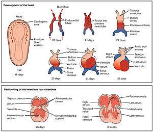
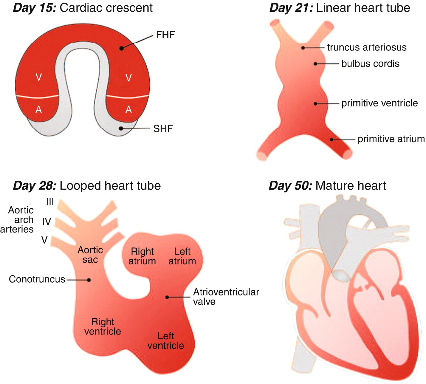
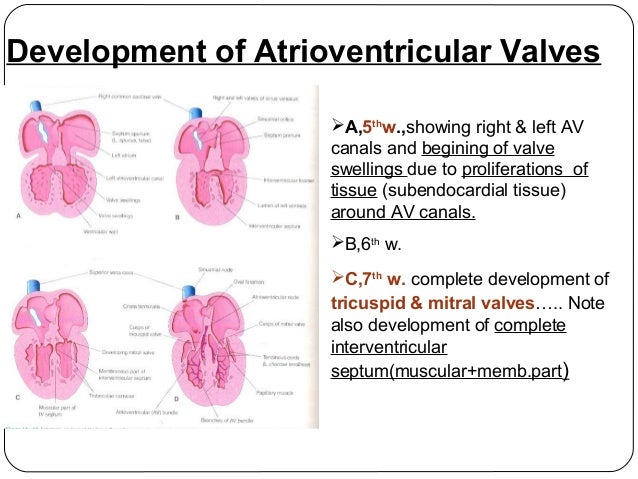
Malfunctioning heart valves inhibit the hearts ability to pump blood and life giving oxygen and nutrients to the cells of the body. These appear at the end of the fourth week at the superior and inferior borders of the atrioventricular canal.

Wt1 Cre Mg Cells Significantly Contribute To The Formation Of The
A malformation is an abnormal connection between the veins and arteries.
Av valve formation. Arteriovenous malformations avms are defects in the blood vessels of the circulatory system. The chordae tendineae which insert into small muscles attached to the ventricle wall. This process begins with the specific processes that contribute to the appearance of the discrete structure and ends when the structural rudiment is recognizable.
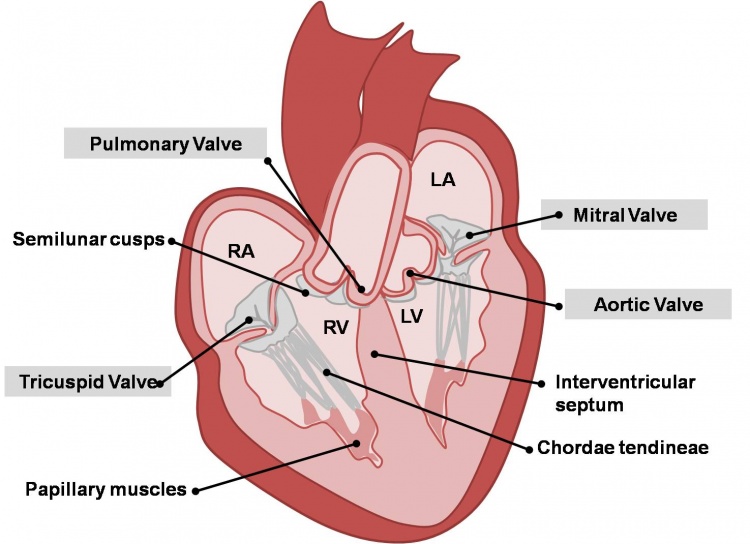
These valves open and close during the cardiac cycle to direct the flow of blood through the heart chambers and out to the rest of the body. During embryonic development the heart is the first organ to function and it forms initially as a primitive tube composed of a myocardial cell layer surrounding an endocardial endothelial cell layer the first indication of valve development during vertebrate embryogenesis is the formation of endocardial cushions in the outflow tract oft and atrioventricular av. The left av valve has anterior and posterior leaflets and is termed the bicuspid or mitral valve.
Origin and development the formation of the two atrioventricular valves the valves between atria and ventricles which regulate the direction of blood flow through the heart. The heart has two kinds of valves atrioventricular and semilunar valves. This vascular anomaly is widely known because of its occurrence in the central nervous system usually cerebral avm but can appear in any location.
Valve formation in the atrioventricular canal and truncus arteriosus there are two swellings of mesenchyme in the wall of the atrioventricular canal that form the endocardial cushions. Arteriovenous malformation is an abnormal connection between arteries and veins bypassing the capillary system. The valve leaflets are attached to the ventricular walls by thin fibrous chords.
A brain arteriovenous malformation avm is a tangle of abnormal blood vessels connecting arteries and veins in the brain. A brain avm disrupts this vital process. In human cardiovascular system.
The right av valve has a third small septal cusp and thus is called the tricuspid valve. Heart valves are formed from elastic connective tissue which provides the flexibility needed to open and close properly. The arteries are responsible for taking oxygen rich blood from the heart to the brain.
This interferes with your. Veins carry the oxygen depleted blood back to the lungs and heart. The av valves begin to form between the fifth and eighth weeks of development.
The av valves begin to form between the fifth and eighth weeks of development. The developmental process pertaining to the initial formation of the atrioventricular valve from unspecified parts.
The Aortic And Pulmonary Valves Semilunar Valves

Http Www Columbia Edu Itc Hs Medical Humandev 2005 Heart2 Pdf
Heart Development Wikipedia

Formation Of The Tricuspid Valve In The Human Heart Circulation
Heart Embryology Overview Springerlink
11 Development Of The Heart

Partitioning The Heart Mechanisms Of Cardiac Septation And Valve
Intermediate Heart Valves Embryology

Partitioning The Heart Mechanisms Of Cardiac Septation And Valve